Thursday, February 25, 2016

Owl Pellet Lab

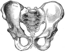
     In this lab, my partner and I dissected an owl pellet. First, we divided the pellet in half and worked on our halves separately. Using forceps and a probe, we picked apart the owl pellet. We found leg bones and some ribs but unfortunately, there was no skull in the pellet. We put together the bones that we found and came to the conclusion that the animal was a shrew. We knew that the animal had to be a rodent of some kind because we found lots of hair in the pellet, meaning the animal couldn't have been a bird. Next, we looked at the differences in bone structure in shrews, moles, and voles. We found two pieces of a pelvis that looked like that of a shrew because of the loops at the ends of the bones. The shape of the femur of the shrew also matched the femur that we found in the pellet. We also found a lower back leg that could have been that of a vole or a shrew; however, we narrowed it down to a shrew's back leg because of the shapes at the ends of the tibia and fibula, as well as the space between the two bones. So by looking at the pelvis, the upper back leg bones, and the lower back leg bones we found, we concluded that the animal was indeed a shrew.


human pelvis
human tibia & fibula
After putting together the shrew skeleton, we could see that it was actually quite similar to a human skeleton. The pelvis of the shrew was like a stretched out version of a human's. The shrew's had sockets where the femur would connect, the same in a human. The ribs we found were also curved in the same way human ribs are. The tibia and the fibula were also similar to a humans because of their shape. Like a human's, the shrew's fibula was very thin and the tibia was very thick. There were also some small differences in the shrew bones in comparison to a human bone. For example, the pelvis of the shrew didn't have a wide ilium; the shrew's ilium was thin and long. The shrew's fibula and the tibia are connected at the bottom and in a human, the bones are separated. The space between the fibula and tibia in a human are not as wide or as curved as the shrew's.


Thursday, January 28, 2016

Unit 5 Reflection


     Unit 5 was about the digestive system, endocrine system, and lymphatic system. We learned about the organs involved in the systems and all their functions. To further understand the digestive system we measured our alimentary canal and made it with string and ribbon. We learned about fuel metabolism and the fed, fasting, and starvation states. We went over how our organs work in different conditions, for example how the brain uses glucose for fuel and the muscles can use glucose, fatty acids, and ketone bodies for fuel. We learned a lot about insulin and glucagon, two hormones that regulate blood glucose levels. A big part of this unit was about diabetes, which I didn't know much about at first. Now I have a greater understanding of the way insulin works in our bodies and how diabetes can be caused by so many different factors such as genetics, stress, and physical inactivity.


     I enjoyed learning about the body systems because they were pretty straightforward. I struggled a little with understanding fuel metabolism because there were so many steps and hormones that played a part in the whole process. I wanted to learn more about metabolism and lactose intolerance and I got the chance to when we had researched and discussed articles in class. So far I have been keeping up with my New Year's goals. I still need to work on getting 7-8 hours of sleep everyday but I have cut down on my screen time.

Wednesday, January 6, 2016

Digestive System Lab

1. In this lab, I measured my digestive system out with ribbon and string. I learned that my digestive system stretched almost all the way across the classroom. I liked this lab because it really put into perspective how long our digestive system is and made me think about how the alimentary canal can fit into the human body.

2. The length of my digestive system is almost 5x longer than my height. My digestive system fits inside my abdomen because the small and large intestines are folded into a small

3. I think it takes a few hours for food to move through the entire digestive system. According to Mayo Clinic, however, food can take 24 to 72 hours to digest. This time varies based on age, amount of physical activity, and the type of food being eaten. For example, the body digests fiber very quickly compared to fruits and vegetables.

4. Digestion is breaking down food into smaller pieces. It begins in the stomach and occurs in the small intestine. Absorption is taking in nutrients and water, which occurs in the small and large intestine.

5. I want to learn more about the enzymes that break down the food and about the structures of the organs of the digestive system.

Tuesday, January 5, 2016

New Year Goals

      For 2016, one of my SMART goals is to cut down the time I spend watching TV or using social media. Spending time on my phone leads to procrastination and staring at a screen for hours isn't healthy. I will put my phone away while doing homework to steer clear of its distractions. Not getting work done efficiently has made me lose sleep as well. I'm constantly tired and can't focus during school. This is why my second SMART goal is to get more sleep. I will make sure to get at least 7-8 hours of sleep per night by making sure I finish my homework before 11 pm. Hopefully this will improve my grades as well. Both of these relate back to anatomy and physiology, especially getting some sleep.

Thursday, November 12, 2015

Heart Dissection Lab

     1. The purpose of the pericardium is to protect the heart.
     2. Arteries take blood away from the heart and veins bring blood to the heart. Valves have thinner walls and have one-way valves to prevent backflow.
     3. The auricle inflates when blood pumps into the heart and increases the volume of the atrium.
     4. There is more fat at the top of the heart near the atrium. The ventricles have thicker walls than the atria walls.
     6.
     7. The chordate tendinae and the papillary muscles are important to heart function because they
     8.

     9. The semi-lunar valves prevent blood from the atrium from entering the heart again.
    10. Valve disease on the right side of the heart results in swelling in the feet and ankles because the blood returns to that area of the body. Since the tricuspid valves stops working, blood would go back through the inferior vena cava and back to the lower part of the body.
          b) If valve disease occured on the left side of the heart, it would mess up the blood flow between the lungs and the left atrium. This would interrupt the flow of oxygenated blood to the rest of the heart.
     11.

     12. The left side of the heart deals with oxygenated blood and the right side deals with deoxygenated blood.

13.

Unit 3 Reflection

      Unit 3 was about the cardiovascular system and cardiovascular health. The cardiovascular system transports materials like oxygen and glucose to cells and removes waste materials from cells. It is made up of the heart, or pump, a network of tubes, and blood. Cardiovascular health is preventing heart diseases and strokes. Heart attacks are occur when blood supply to the myocardium is reduced or completely stopped. This results in damage to heart cells. A stroke occurs when an artery which supplies the brain becomes blocked. Both strokes and heart attacks can be caused by atherosclerosis, chronic inflammation in blood vessels. To prevent atherosclerosis or any cardiovascular disease, one should control blood pressure, cholesterol,  and weight. One should eat healthfully, exercise, and manage stress as well. I would want to learn more about the treatments of heart attacks and stroke. I still have questions about the ways each treatment is different and how doctors decide which treatment they would use for their patients.

        I enjoyed this unit and it was because of the labs. For example, doing the sheep heart dissection really put everything into perspective. It was so different from the diagrams and models we had looked at in class. Having to find the parts of the heart from memory and from definitions was challenging but I learned a lot from it. Towards the end of the unit, however, I started to slack off on reviewing my lecture notes. I think this set me back a bit because I didn't remember that material very well and a few days before the test I was trying to cram all that information into my head. For the rest of the semester I will make sure I fully understand the material to be able to apply it to real life situations.