Unit 6 was all about the skeletal system. First, we learned about the different types of bones, what bones are made of, and how they repair themselves. The axial bones are the bones that make up our foundation and protect our organs. The appendicular bones help us with movement. Bones are classified by their shape: long, short, flat, and irregular. The two types of bone tissue of bone are compact and spongy. The cells that are responsible for bone remodeling are osteocytes, osteoblasts, and osteoclasts. Osteoblasts are bone forming cells and osteoclasts are bone destroying cells. Next, we learned about the disorders of the skeletal system like arthritis. Osteoporosis and rickets are caused by a deficiency in vitamins and minerals. We also learned about kyphosis, lordosis, and scoliosis which are all diseases relating to an abnormal curvature of the spine. Lastly, we learned the names of all the bones and the different kinds of joints.
I want to learn more about the cures for bone diseases, if any. Do you have to live with the disease forever or are some of the diseases reversible? For example, osteoporosis is by loss of minerals in the bones, making them more brittle. Does this mean a person with osteoporosis just needs to have more minerals in their diet or is there something more? I would also like to learn more about the similarities and differences between human bones and other animal bones because of the owl pellet lab. So far this semester has been going well and I've been keeping up with my New Years goals.
My blog is about my experiences in this anatomy and physiology class. It serves as a journal to record my growth throughout the year.
Saturday, February 27, 2016
Thursday, February 25, 2016
Owl Pellet Lab
 In this lab, my partner and I dissected an owl pellet. First, we divided the pellet in half and worked on our halves separately. Using forceps and a probe, we picked apart the owl pellet. We found leg bones and some ribs but unfortunately, there was no skull in the pellet. We put together the bones that we found and came to the conclusion that the animal was a shrew. We knew that the animal had to be a rodent of some kind because we found lots of hair in the pellet, meaning the animal couldn't have been a bird. Next, we looked at the differences in bone structure in shrews, moles, and voles. We found two pieces of a pelvis that looked like that of a shrew because of the loops at the ends of the bones. The shape of the femur of the shrew also matched the femur that we found in the pellet. We also found a lower back leg that could have been that of a vole or a shrew; however, we narrowed it down to a shrew's back leg because of the shapes at the ends of the tibia and fibula, as well as the space between the two bones. So by looking at the pelvis, the upper back leg bones, and the lower back leg bones we found, we concluded that the animal was indeed a shrew.
 |
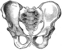
| human pelvis |
 |
| human tibia & fibula |

Subscribe to:
Comments (Atom)