Tuesday, May 31, 2016

20 Time Reflection


      I can't believe we finished presenting our TED Talk! The semester went by so fast and I'm proud of the presentation Alyssa and I gave! We achieved our goal of informing people about the drought through our TED talk. We talked about the causes of the drought relating to agriculture, specifically the types of crops that are grown in California and factory farming. We also found solutions to the drought like desalination, drip irrigation, and recycling water. We didn't get to our final project: filmed interviews of people in our community asking them what they thought about the drought. This way we could find out what the public already knew about the drought. We didn't want to ramble off about things that everyone already knew about during our TED talk. We also would have liked to talk to some officials, people who study the drought every day and how it's affecting our community. The most successful part of our project was the survey we put out. We got almost 50 responses and it helped us find the focus of our project.
      Our TED talk went well! I did talk quickly because I was a bit nervous. There was just so much information Alyssa and I wanted to include in our project but we had to stick to the time limit. While prepping for the presentation, we had gone on talking for a really long time. We were really passionate about the subjects and we wanted to share everything we had learned with the class. It was hard to cut down on the info. In all I learned that you really need to plan out a timeline for big projects. I think the reason we sort of fell behind in was because we only planned what we needed to do for a week, not for multiple weeks. We also didn't find the focus for our project very early on, since the drought has so many different aspects. So time management will be something that I continue to work on. I also learned a lot about teamwork. This 20 Time project was a great experience especially because I got to work on something that I'm passionate about and will help the environment! 

Wednesday, May 11, 2016

Reflexes Lab Analysis

      In this lab we tested our reflexes! First, we tested the photopupillary reflex. The smooth muscles in our eyes control the size of our pupils. If an intense light is shined into the eye, the pupil's size decreases keeping all the light from entering and damaging the eye. This reflex protects the photoreceptors in our eyes.

      Next, we tested the knee jerk reflex. At first, we didn't get any results. But after multiple tries and help from other people, my leg moved involuntarily. It was a very weird feeling. My patella tendon had flexed. I learned that this reflex is what helps us keep balance while walking and running. After doing a few squats, we tested the reflex again. This time my leg didn't move. This is probably because my thigh muscles were fatigued from the squats. There was not enough ATP in my leg for the muscle to contract.

      We also tested our blink reflexes. Even with a shield to cover our eyes from the cotton ball, both of us blinked when we saw it coming towards us. This reflex protects objects from flying into our eyes and damaging them. Then, we tested the plantar reflex. When dragging a pen up the foot, the toes flex and move close together. People with Multiple Sclerosis are more likely to show Babinski's sign. If there is nerve damage in the foot, the toes spread apart instead of together.

     Lastly, we tested our response time to something that we see. It took me an average time of 0.20 seconds to catch a falling yardstick. While on my phone, it took me an average time of 0.29 seconds to catch the falling yardstick. It was hard to focus on catching the yardstick without looking at it. This is important for drivers to understand. The brain is not very good at multitasking, especially when it can't see one of the tasks it is doing. My response time was almost 0.10 seconds slower while texting. Those 0.10 seconds could mean the difference between life and death on the road.
Here are the average response times of our class!

Thursday, May 5, 2016

Brain Map

Cerebral Cortex
  • What do the frontal lobes do?
    • The frontal lobes are the command center of the brain. They control personalities, problem-solving, memory, language, and judgement.
  • What is the relationship between selective attention and learning?
    • Selective attention makes your memory capacity greater so you can prioritize more important information above little-used information. 
  • What is the last part of your brain to develop and what can you do to prevent it from deteriorating? 
    • The frontal lobe is the last part of the brain to develop. To prevent the frontal lobe from deteriorating, always be engaged with your environment and transform information instead of just memorizing it. 
  • What does the neo cortex do?
    • The neo cortex helps us navigate our bodies. It controls our senses, spatial awareness, and motor skills.
  • What is the role of the pre frontal cortex?
    • The pre frontal cortex controls our personalities and our behavior in social situations. 
  • What do we know about the pre frontal cortex’s relationship with multitasking?
    • It prevents us from doing more than two big tasks at the same time. We can't actually multitask; our brain jumps back and forth between tasks, meaning we do both tasks inadequately.  
  • Which part of the brain is associated with speech and language development?  Give an interesting fact about this region.
    • Broca's Area is responsible for speech production and language development. Mirror neurons are found in this part of the brain and they to learn language production and comprehension.
  • Which part of your brain is responsible for thinking the following: “Is it hot in here or is it just me?”
    • The somatosensory cortex responds to touch and interprets temperature.
  • What does your visual cortex do for you?
    • The visual cortex differentiates colors and faces.
  • State three interesting or significant facts about your occipital lobe.
    • collects and catagorizes visual information to be stored in other parts of the brain
    • deals with imagination and processing memories
    • visualizing doing a task multiple times will help you perform that task better
  • What would happen if your temporal lobes were damaged?
    • If your temporal lobes were damaged, you wouldn't have a long term memory so you would forget things like faces and the alphabet.
  • What is your “fast brain” and what does it do?
    • The eye fields control eye movement and help the brain process movement quickly.

Neuron
  • State 3 things that you could do that would influence your synapses, and have a positive affect on your life and health.
    • Active learning, sleep, and exercise would influence your synapses. 
  • What is the relationship between multi-sensory or multi-modal learning and your dendrites?
    • By engaging multiple parts of the brain in storing information, the brain will convert the information from memorized to learned.
  • How does “big picture thinking” and mnemonics affect dendrites and/or learning?
    • "Big picture thinking" helps one to fully comprehend a subject. Mnemonics help us remember subjects based on personal or relatable information. 
  • Describe a neurotransmitter that you feel is very important.  Justify your reasoning.
    • Glutamate is a very important neurotransmitter because it helps with long term memory. It is also affects the way we learn and strengthens our synapses. 


Limbic System
  • What does the corpus callosum do?
    • The corpus callosum communicates between the left and right hemispheres of the brain. It is responsible for motor, cognitive, and sensory functions between the hemispheres.
  • What is the relationship between music and the corpus callosum
    • Studies have shown that musical practice can strengthen the communication between the two hemispheres of the brain. 
  • Why is the thalamus important?
    • The thalamus relays information from the senses to the cortex. It also processes some information.


I learned more about the way all the parts of the brain work together. I also realized that multiple parts of the brain had similar functions. I think that's why if one part of the brain is damaged, it can still function normally, because the other parts of the brain take over for it. I learned about different neurotransmitters like oxytocin, glutamate, and dopamine. I learned the parts of the limbic system that I didn't really know about before like the basal ganglia and amygdala. 

Tuesday, May 3, 2016

Sheep Brain Dissection

We dissected a brain!  First, we took off some of the meninges. Even though the two hemispheres of the brain are only connected by the corpus callosum, it wasn't easy to pull both sides apart because of the meninges. I also noticed that in some of the parts where where the meninges were removed, the brain had lost it's structure. We marked the parts of the brain with pins. Then we cut the brain into the right and left hemispheres. At first, it was hard to identify some of the parts like the optic nerve, even though they were clearly seen in the book. Lastly, we cut the cerebrum to see the center of it. It was very easy to tell white matter from gray matter.


Thursday, April 28, 2016

Sheep Eye Dissection

Here's a picture of the eye before we dissected it! The oval shaped structure in the center is the cornea, which helps focus the light entering the eye. Surrounding the cornea is the sclera, also known as the white of the eye. The sclera is a tough outer coat that protects the eye. It was very hard to cut into it. On top of the sclera is fatty tissue and external eye muscle remnants that we cut off later.
Cornea and sclera seen from the outside of the eye.

Cornea and sclera seen from the inside of the eye.


The small bump on the top of the eye is the optic nerve which sends the visual information from the retina to the brain. 
Here is the eye cut into two hemispheres. On the left, there is the retina and tapetum lucidum. The retina, bunched up at the top on the left, contains the photoreceptors for vision. The blue part under the retina is the tapetum lucidum. It's part of the choroid layer and reflects light onto the retina. The choroid is the black layer that  nourishes the back of the eye. The tapetum lucidum isn't found in humans, but it is in the eyes of many nocturnal animals to help them see at night. The ciliary body is in the right side of the eye. It is made up of muscles and controls and shapes the lens.

The round structure at the bottom of the picture is the lens. The lens changes shape to focus light on the retina. I thought that it would be squishy because it changes shape; however, the lens was hard like a marble. The liquid, which had the consistency of a gel, around the lens is the vitreous humor. It fills the central cavity of the eye. The suspensory ligament can be seen around the lens and connects the lens to the ciliary body.

This is the iris, or the colored portion of the eye. The hole in the center is the pupil. This is where light passes through into the eye.


Thursday, April 14, 2016

The Clay Brain

     Our new unit is about the brain, so we made one out of clay in class! After looking through the textbook we started to outline the shape of the brain. We used different colored clay for each part of the brain and then labelled the parts. We shaped the bigger parts of the brain first, like the frontal lobe and temporal lobe. Then we added the smaller parts of the brain like the transverse fissure and central sulcus in between the lobes. This activity helped me learn the names of parts of the brain and how they all are connected.

Wednesday, April 13, 2016

Missing Parts of the Brain?

     This article is about a woman was able to survive for 24 years without a cerebellum. The cerebellum is essential for motor control, muscle memory, and speech. It modifies the motor commands before sending them to the muscles to make sure the command is accurate. The cerebellum also has about half of the total neurons in the brain. This would explain why the woman had problems keeping balance while walking and felt dizzy. Missing part of the brain is a rare case; cases like these reveal how the brain is able adapt and continue to function even without a large part of it.
     The lateral cerebral sulcus separates the frontal and parietal lobes from the temporal lobe. The sulci are the grooves in the brain. These folds help the brain take in more glucose and oxygen and help the brain fit into our skull. The more grooves an organism's brain has, the more complex functions the organism can perform. An absence of these grooves is known as lissencephaly, which causes developmental delays and brain malformation. Polymicrogyria is a condition where there are too many folds in the brain which can cause neurological problems like seizures, delayed development, and muscle weakness.
   

Sunday, March 27, 2016

Unit 7 Reflection

 This unit was all about muscles! Some of the main themes of the unit were basic anatomy of the muscle, how muscles contract and relax, and muscle health. First, we learned about directional terms and joint movements through a dance we made up. The dance was a really fun and easy way to memorize the many movements. Next, we learned about muscles from a more anatomical point of view. Our muscle contraction video was a creative way to show we really understood what is happening in the muscle when it contracts. We reviewed the three types of muscle tissue: skeletal, smooth, and cardiac. We learned about connective tissue, which was similar to the connective tissue around the heart. We also went over how the muscles work with one another to move parts of the body, like bending the elbow or flexing the foot. We also learned the names of the major muscles in the body and how they're named based on their size, shape, location, and action. The chicken dissection helped me review the muscles and their names. Lastly, we learned about the ways lifestyle affects muscle health. For example, we learned the difference between the muscles of a sprinter and a long distance runner. We also went over the effects of steroids on the body.
                                                                                Here's our muscle contraction video!


Thursday, March 24, 2016

Performance Enhancement Ad: High Protein Diets

      Performance enhancing substances have more risks than benefits. The stereotype that men have to be super muscular and women have to be super thin is definitely a very dangerous stereotype that is portrayed in the media. Athletes also have a lot of peer and parental pressure to be the best they can be, leading them to believe that performance enhancement substances are the only way to achieve their goals. Even though, performance enhancements can boost athletic performance, build muscle, and help with weight loss, the long term effects can be very harmful. For example, a high protein diet may help you get a six-pack but it will also increase LDL levels and build up toxic ketones in the body. Caffeine seems harmless but it can cause dehydration, heat stroke, insomnia, and can lead to addiction. In all, I think the best way to enhance performance is to work hard.

Monday, March 21, 2016

Chicken Dissection Analysis

     This week we dissected a chicken! I have a much better understanding of the muscles now; it was really helpful to see the muscles attached to the bones and the tendons in front of me instead of on a paper. Signals from our brain are sent to muscles so that we can move. Tendons connect the muscles to the bone and help the muscles pull on the bones. While stretching, muscles move the bones by working together. For example, we are able to bend our elbow because the biceps and the triceps work together. When you bend your elbow, the bicep muscle contracts and the triceps muscle is stretched out. A big difference in the tendon of the insertion and the tendon of the origin is where they are located. Insertion is the movable end of the muscle, while origin is the immovable end of the muscle. The tendon of the insertion has to be more flexible than the tendon at the origin.
      Chicken muscles are pretty similar to human muscles function wise. For example, the wing muscles of the chicken was similar to the muscles in a human arm. In both the chicken and human, the biceps brachii flexes the elbow joint. The deltoid of the chicken also looked similar to a human deltoid in size and shape. There were also muscles that were very different in shape, like the trapezius. The chicken's trapezius ran from the backbone to the shoulder of the bird. In a human, the trapezius is split into two parts: one up the neck and one along the back. The whole front part the chicken was the pectoralis major. There was no rectus abdominus or external obliques in the chicken as there are in humans.

Here are the pictures we took!
The sartorius is the longest muscle in the body. It is responsible for lateral hip rotation and flexion of the thigh and knee,
The triceps humeralis extends the wing (birds) or arm. 
The trapezius extends the head. It also elevates and depresses the scapula.
The pectoralis minor pulls the shoulder down and forward.

The deltoid is responsible for the abduction, flexion, and extension of the arm.

The biceps femoris is part of the hamstring group. It flexes the lower leg.

The biceps brachii flexes the elbow joint. 

The iliotibialis extends the thigh and flexes the leg.

The latissimus dorsi extends, adducts, and rotates the arm medially.

The semitendinosus is anterior and medial to the inner thigh. It works with the semimembranosus to extend the thigh.

The gastrocnemius is the primary muscle of the posterior and medial sides of the drumstick.

The brachioradialis flexes the forearm at the elbow. It is responsible for pronation and supination.

The pectoralis major pull the wings of a bird ventrally so they can fly. In a human, the muscle flexes, adducts, and rotates the arm.


The peroneus longus is responsible for the extension of the foot. 

The flexor carpi ulnaris is responsible for the flexion of the hand.
The tibialis anterior runs along the lateral side of the shin bone and flexes the foot. 
We found tendons!

Monday, March 14, 2016

What Happens When You Stretch?

     This article explained what stretching is and the effects of stretching on muscles. I learned why stretching caused flexibility from an anatomical perspective. I learned that connective tissue plays a big role in stretching; it's helps when a muscle is stretched further than normal. Stretching realigns fibers and fixed scarred tissue, which explains why stretching helps with soreness. The article also explained reciprocal inhibition and how the muscles contract and relax to cause a desired motion. I thought the most interesting part of the article was how muscle spindles work. Sudden changes in muscle length can cause strong muscle contractions so muscle spindles help maintain muscle tone and prevent injuries. You could even train the stretch reflex of the muscles, increasing flexibility.

-"The more fibers stretched, the more length developed by the muscle for a given stretch." (1) - this gave me a better understanding of flexibility of the muscle. I knew that stretching got rid of soreness, but I did not know how that correlated with muscle fibers.

-"as you hold the muscle in a stretched position, the muscle spindle habituates and reduces its signaling" (3) - this explained why people must stretch every day in order to be able to do a split or just touch their toes. After a while, the muscle is able to stretch further and further because of the reduction of signaling.

-"When you stretch your calf, you want to contract the shin muscles by flexing your foot. However, the hamstrings use the calf as a synergist so you want to also relax the hamstrings by contracting the quadricep" (2) - I found it interesting how simply flexing the foot could affect multiple muscles as far up as the quads and hamstrings. It's like a chain reaction; each muscle lifts up another muscle.

Saturday, February 27, 2016

Unit 6 Reflection

      Unit 6 was all about the skeletal system. First, we learned about the different types of bones, what bones are made of, and how they repair themselves. The axial bones are the bones that make up our foundation and protect our organs. The appendicular bones help us with movement. Bones are classified by their shape: long, short, flat, and irregular. The two types of bone tissue of bone are compact and spongy. The cells that are responsible for bone remodeling are osteocytes, osteoblasts, and osteoclasts. Osteoblasts are bone forming cells and osteoclasts are bone destroying cells. Next, we learned about the disorders of the skeletal system like arthritis. Osteoporosis and rickets are caused by a deficiency in vitamins and minerals. We also learned about kyphosis, lordosis, and scoliosis which are all diseases relating to an abnormal curvature of the spine. Lastly, we learned the names of all the bones and the different kinds of joints.

      I want to learn more about the cures for bone diseases, if any. Do you have to live with the disease forever or are some of the diseases reversible? For example, osteoporosis is by loss of minerals in the bones, making them more brittle. Does this mean a person with osteoporosis just needs to have more minerals in their diet or is there something more? I would also like to learn more about the similarities and differences between human bones and other animal bones because of the owl pellet lab. So far this semester has been going well and I've been keeping up with my New Years goals.

Thursday, February 25, 2016

Owl Pellet Lab

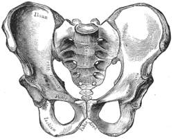
     In this lab, my partner and I dissected an owl pellet. First, we divided the pellet in half and worked on our halves separately. Using forceps and a probe, we picked apart the owl pellet. We found leg bones and some ribs but unfortunately, there was no skull in the pellet. We put together the bones that we found and came to the conclusion that the animal was a shrew. We knew that the animal had to be a rodent of some kind because we found lots of hair in the pellet, meaning the animal couldn't have been a bird. Next, we looked at the differences in bone structure in shrews, moles, and voles. We found two pieces of a pelvis that looked like that of a shrew because of the loops at the ends of the bones. The shape of the femur of the shrew also matched the femur that we found in the pellet. We also found a lower back leg that could have been that of a vole or a shrew; however, we narrowed it down to a shrew's back leg because of the shapes at the ends of the tibia and fibula, as well as the space between the two bones. So by looking at the pelvis, the upper back leg bones, and the lower back leg bones we found, we concluded that the animal was indeed a shrew.


human pelvis
human tibia & fibula
After putting together the shrew skeleton, we could see that it was actually quite similar to a human skeleton. The pelvis of the shrew was like a stretched out version of a human's. The shrew's had sockets where the femur would connect, the same in a human. The ribs we found were also curved in the same way human ribs are. The tibia and the fibula were also similar to a humans because of their shape. Like a human's, the shrew's fibula was very thin and the tibia was very thick. There were also some small differences in the shrew bones in comparison to a human bone. For example, the pelvis of the shrew didn't have a wide ilium; the shrew's ilium was thin and long. The shrew's fibula and the tibia are connected at the bottom and in a human, the bones are separated. The space between the fibula and tibia in a human are not as wide or as curved as the shrew's.


Thursday, January 28, 2016

Unit 5 Reflection


     Unit 5 was about the digestive system, endocrine system, and lymphatic system. We learned about the organs involved in the systems and all their functions. To further understand the digestive system we measured our alimentary canal and made it with string and ribbon. We learned about fuel metabolism and the fed, fasting, and starvation states. We went over how our organs work in different conditions, for example how the brain uses glucose for fuel and the muscles can use glucose, fatty acids, and ketone bodies for fuel. We learned a lot about insulin and glucagon, two hormones that regulate blood glucose levels. A big part of this unit was about diabetes, which I didn't know much about at first. Now I have a greater understanding of the way insulin works in our bodies and how diabetes can be caused by so many different factors such as genetics, stress, and physical inactivity.


     I enjoyed learning about the body systems because they were pretty straightforward. I struggled a little with understanding fuel metabolism because there were so many steps and hormones that played a part in the whole process. I wanted to learn more about metabolism and lactose intolerance and I got the chance to when we had researched and discussed articles in class. So far I have been keeping up with my New Year's goals. I still need to work on getting 7-8 hours of sleep everyday but I have cut down on my screen time.

Wednesday, January 6, 2016

Digestive System Lab

1. In this lab, I measured my digestive system out with ribbon and string. I learned that my digestive system stretched almost all the way across the classroom. I liked this lab because it really put into perspective how long our digestive system is and made me think about how the alimentary canal can fit into the human body.

2. The length of my digestive system is almost 5x longer than my height. My digestive system fits inside my abdomen because the small and large intestines are folded into a small

3. I think it takes a few hours for food to move through the entire digestive system. According to Mayo Clinic, however, food can take 24 to 72 hours to digest. This time varies based on age, amount of physical activity, and the type of food being eaten. For example, the body digests fiber very quickly compared to fruits and vegetables.

4. Digestion is breaking down food into smaller pieces. It begins in the stomach and occurs in the small intestine. Absorption is taking in nutrients and water, which occurs in the small and large intestine.

5. I want to learn more about the enzymes that break down the food and about the structures of the organs of the digestive system.

Tuesday, January 5, 2016

New Year Goals

      For 2016, one of my SMART goals is to cut down the time I spend watching TV or using social media. Spending time on my phone leads to procrastination and staring at a screen for hours isn't healthy. I will put my phone away while doing homework to steer clear of its distractions. Not getting work done efficiently has made me lose sleep as well. I'm constantly tired and can't focus during school. This is why my second SMART goal is to get more sleep. I will make sure to get at least 7-8 hours of sleep per night by making sure I finish my homework before 11 pm. Hopefully this will improve my grades as well. Both of these relate back to anatomy and physiology, especially getting some sleep.